
Spinal Cord(상행로)
Connections of the dorsal horn and dispersion of the dorsal roots
A. Nuclear grouping of the dorsal horns. The dorsal horns contain several nuclei or laminae that are directly in the path of the entering dorsal root axons. These include the:
- Dorsomaginal nucleus
- Substantia gelatinosa
- Nucleus proprius
- Nucleus dorsalis of Clarke
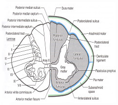
- The more ventral parts if the ventral horn contain laminae VIII and IX of rexed
- It receives supramaginal and dorsal root fibers
- It connect with ventral horn motorneurons
- Somatic nuclei. Lamina IX contains the medial and lateral motor cell columns
- The medial motor cell column innervates the axial muscles
- The lateral motor cell column innervates the appendicular muscles
Motorneurons of the ventral horn
- Motorneurons of the ventral horn belong to one of two types:
- a. Large, or alpha, motorneurons
- b. Small, gamma, motorneurons
- Characteristics of alpha motorneurons
- a. In general, large perikarya have large dendritic trees, large axons, or both
- Ventral motorneurons
- GSE neurons in the spinal cord and brainstem
- Special visceral efferent neurons(SVE) 0f the brainstem
- Lamina IX of the Rexed
- Lower motorneurons(LMNs)
- Final common pathway
- a. In general, large perikarya have large dendritic trees, large axons, or both
- Gamma motorneurons
- a. The smaller gamma motorneurons of the ventral horn send gamma-sized axons into the peripheral nerves
- b. The axons of gamma motorneurons end on specialized skeletal muscle fibers contained in muscle spindle
Concept of motor units
- a. Definition. A motor unit is an alpha motorneuron, its axon, and all of the muscle fibers it innervates
- b. A motor unit is the smallest unit of behavior because the neuron responds on all-or-none principle, the axon conducts on an all-or- none, and muscle fiber responds all-or-none
Motor unit fasciculation
disease may destabilize the membrane of the alpha motorneuron or its axon, causing impulses to arise spontaneously, rather than in response to normal synaptic excitation. Thus motor unit is the anatomical unit, functional, pathologic, and regenerative unit of the neuromuscular system.
Renshaw cell
- The alpha motorneuron send a collateral axon to an interneuron, the Renshaw cell, which sends an inhibitory synapse back to the alpha motorneurons
- Physiological or pathological changes in the balance of excitatory and inhibitory impulses playing on the Renshaw cells and alpha motorneurons may cause the MSRs to be hyperactive or hypoactive, which can be detected when the clinical elicits the MSRs.
ASCENDING PATHWAYS OF THE SPINAL WHITE METTER
A. Features of the ascending pathways
- Ascending pathways run in each of the three columns of white matter, the dorsal, lateral, and ventral
- Ascending long pathways all transmit information derived from sensory
- receptors and destined to:
- a. Produce conscious sensation, somatic or visceral
- b. Mediate reflexes, somatic or visceral
- c. Guide the motor centers in the cerebellum, brainstem, diencephalon, basal ganglia, and cortex
Conscious sensation
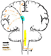
Ascending somatosensory pathways that mediate conscious sensation include the :
- a. Lateral spinothalamic tract (pain and temperature)-Neospinothalamic tract
- b. Ventral spinothalamic tract ( touch)-Paleospinothalamic tract
- c. Fasciculus gracilis and cuneatus of the dorsal column (discriminative modalities: touch, form, texture, position sense)
Unconscious sensation
Ascending somatosensory pathways that mediate unconscious sensory function Include the :
- a. Spinocerebellar pathways provide information to the cerebellum for the coordination of volitional movements
- b. Spinoreticular pathways provide information to the brainsrem, tegmentum and reticular formation to mediate somatic and vesceral reflexes
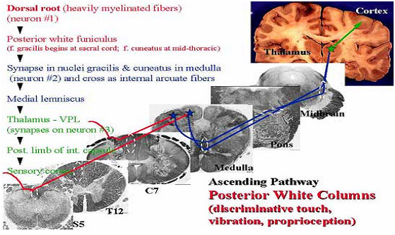
1. Posterior White Column-Medial Lemniscal Pathway
- Modality : Discriminative Touch Sensation (include Vibration) and Conscious Proprioception (Position Sensation, Kinesthesia)
- Receptor : Most receptors except free nerve endings
- Ist Neuron : Dorsal Root Ganglion (Spinal Ganglion) Posterior Root - Posterior White Column
- 2nd Neuron : Dorsal Column Nuclei (Nucleus Gracilis et Cuneatus) Internal Arcuate Fiber - Lemniscal Decussation - Medial Lemniscus
- 3rd Neuron : Thalamus (VPLc) Internal Capsule ----- Corona Radiata
- Termination : Primary Somesthetic Area (S I)
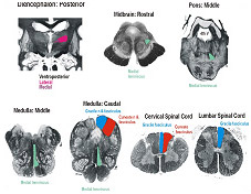
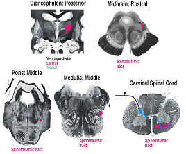
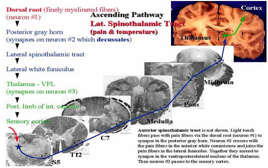
2. Spinothalamic Tract
- Modality: Pain & Temperature Sensation, Light Touch
- Receptor: Free Nerve Ending
- Ist Neuron : Dorsal Root Ganglion (Spinal Ganglion) Posterior Root
- 2nd Neuron : Dorsal Horn (Lamina I, IV, V) Spinothalamic Tract - (Spinal Lemniscus)
- 3rd Neuron : Thalamus (VPLc, CL & POm) Internal Capsule ----- Corona Radiata
- Termination : Primary Somesthetic Area (S I) & Diffuse Widespread Cortical Region
Ipsilateral loss of discriminative touch sensation and conscious proprioception below the level of lesion
Contralateral loss of pain and temperature sensation below the level of lesion
| Fast Pain | Slow Pain |
|---|---|
| Sharp, pricking Group III (A) fiber Short latency Well localized Short duration Less emotional Not blocked by morphine Neospinothalamic tract |
Dull, burning Group IV (C) fiber Slower onset Diffuse Long duration Emotional, autonomic response Blocked by morphine Paleospinothalamic tract |
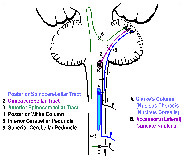
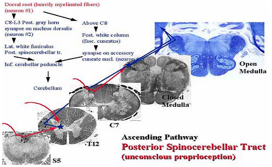
3. Spinocerebellar Tract
- Modality: Unconscious Proprioception
- Receptor: Muscle spindle, Golgi tendon organ
- Ist Neuron :
- Dorsal Root Ganglion (Spinal Ganglion)
- Posterior Root , [Posterior Column]
- 2nd Neuron :
- Clarke's column - Posterior Spinocerebellar Tract
- Accessory Cuneate Nucleus - Cuneocerebellar Tract
- Posterior Horn Anterior - Spinocerebellar Tract
- Termination :
- Cerebellar Cortex
Function of Spinocerebellar Tracts
- Information that travels in the spinocerebellar tracts is not consciously perceived
- Used for unconscious adjustments to movement and posture Because the internal feedback tract
- convey descending motor information to the cerebellum prior to information reaching the motor neurons Because the high-fidelity pathways
- convey information from the muscle spindle, golgi tendon organs and cutaneous mechanoreceptor
The cerebellum obtains information about the movements and postural
Adjustments that followed the command. Thus, cerebellum compare the intended motor output with the actual movement output.