
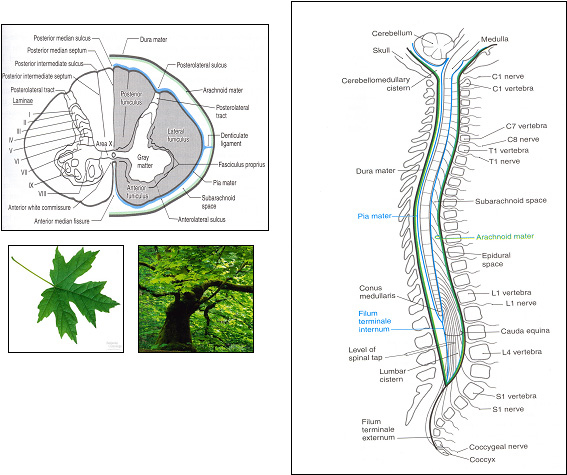
DESCENDING PATHWAYS OF THE SPINAL WHITE METTER
Sources
-
Spinal sources include:
- Short pathways in the ground bundles
- Descending collaterals of dorsal roots in the dorsal columns and dorsolateral tract of Lissauer
-
Supraspinal sources include the cerebral cortex and brain stem
- In the cerebral cortex, only the region around the central sulcus(the sensorimotor cortex) gives rise to clinically significant descending tracts that descend directly into the spinal cord.
- No tracts from the cerebellum, diencephalon, or basal ganglia descend directly into the spinal cord
-
Descending motor Tracts
Upper motor neurons project from supraspinal centers to lower motor neurons(alpha and gamma) and to interneurons in the brain stem and spinal cord
Classified according to where they synapse: medially, laterally, throughout the ventral horn
-
The medial activation system
- controls lower motor neurons that innervate postural and girdle muscles.
-
The lateral activation system
- controls lower motor neurons that innervate distally located muscle used for fine movement
-
Nonspecific activating tracts(throughout the ventral horn)
- contributes to background levels of excitation in the cord and facilitates local reflex arcs.
-
The medial activation system
Medial Activation System - Postural and Gross Movement
Medial activation tracts adjust the activity in the axial and girdle muscle Four tracts from the brain stem and one from the cerebral cortex deliver commands to the medial motor neuron pools
Function of Medial Activation System
- Demonstrated by the reactions generated when a loud noise occurs behind someone.
- The eyes and face turn toward the sound and postural adjustments support the movements, even before the person is consciously aware of the stimulus.
- These coordinated, involuntary reaction occur via circuitry in the brain stem
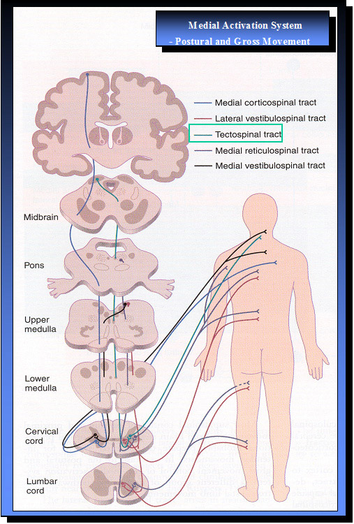
Medial Activation System - Postural and Gross Movement
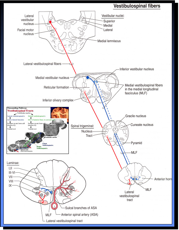
Vestibulospinal tracts
1. Medial Vestibulospinal Tract
- Originate in the medial and inferior vestibular nuclei
- Descending bilaterally into the spinal cord
- Projects only as far as cervical or upper thoracic level
-
Function :
- Influences motor neurons controlling neck and upper back musculature
2. Lateral Vestibulospinal Tract
- Originate in cells of the lateral vestibular nucleus
- Descend ipsilaterally into the spinal cord
- Extend throughout the length of the cord
-
Function :
- facilitate lower motor neurons to extensor while inhibiting lower motor neurons to flexors
- control of posture and balance
These tracts operate constantly to maintain equilibrium in upright positions
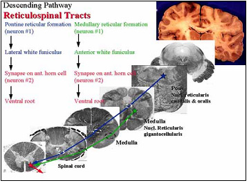
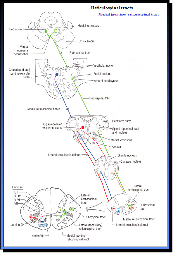
Medial(pontine) Reticulospinal Trace
- Begins in the pontine reticular formation
- Which runs the full length of the spinal cord
- Terminate in anteromedial portions of laminae VII and VIII, where they influence motor neuron supplying paravertebral and limb extensor musculature.
- Function : tend to mediate excitatory effects Stimulation of this tract facilitates ipsilateral lower motor neurons innervating postural muscles and limb extensors.
Tectospinal Tract
- The superior colliculus processes visual, auditory, and somatic information
- The stimulus is auditory
-
Function :
Neural activity in the superior colliculus stimulates neurons that project to the spinal cord in the tectospinal tract, activating lower motor neurons in the cervical spinal cord to reflexively turn the head toward the sound
Quickly turning the head destabilizes a person unless other autonomic responses compensate for the change in
weight destribution.
Loss of balance is prevented by muscle whose motor meurons are activated by neurons in the pontine reticular
formation and vestibular uncleus
Medial Corticospinal Tract
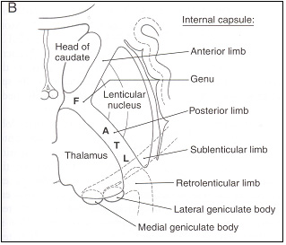
The direct connection from the cerebral cortex to the spinal cord, the medial corticospinal tract, descends from the
cortex through the internal capsule and the anterior brain stem.
The medial corticospinal tract ends in the thoracic cord and thus
assists in control of neck, shoulder, and trunk muscles.
Function of Medial Activation System
- Demonstrated by the reactions generated when a loud noise occurs behind someone.
- The eyes and face turn toward the sound and postural adjustments support the movements, even before the person is consciously aware of the stimulus.
- These coordinated, involuntary reaction occur via circuitry in the brain stem
Lateral Activation System - Limb and Fine Movement
Three tracts controlling distal limb movements descend in the lateral spinal cord and synapse with laterally located motor neuronal pools in the ventral horn
Function of Lateral Activation System
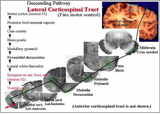
Lateral Corticospinal Tract
Originate in deep portions of layer V of the cerebral cortex (called Betz cells)
Corticospinal neurons are found primarily in six cortical location
-
Function : Control of skilled movement, and modulation of sensory activity
The unique contribution of lateral corticospinal neurons is fractionation of movement.- Fractionation : is the ability to activate individual muscles independently of other muscle. is essential for normal movement of hands, enablings us to tie knots, press individual piano or typewriter keys, and pick up small objects.
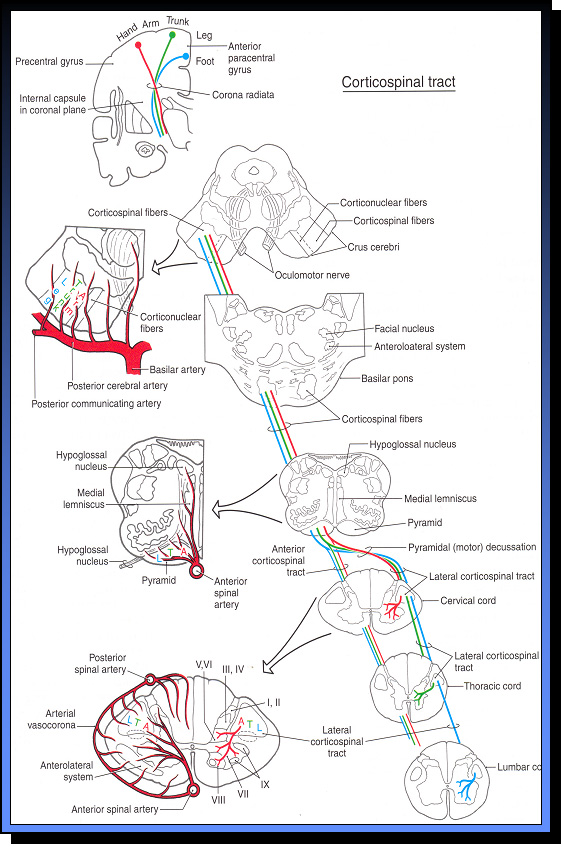
Lateral Corticospinal Tract
- Origin: Cerebral Cortex
- Brodmann Area 4 (Primary Motor Area, M I)
- Brodmann Area 6 (Premotor Area, PM )
- Brodmann Area 3,1,2 (Primary Somesthetic Area, S I)
-
Brodmann Area 5 (Anterior Portion of Sup. Parietal Lobule)
- Corona Radiata
- lnternal Capsule, Posterior Limb
- Crus Cerebri, Middle Portion
- Longitudinal Pontine Fiber
- Pyramid - pyramidal decussation
- Corticospinal Tract - Lateral and Anterior
- Termination: Spinal Gray (Rexed IV-IX)
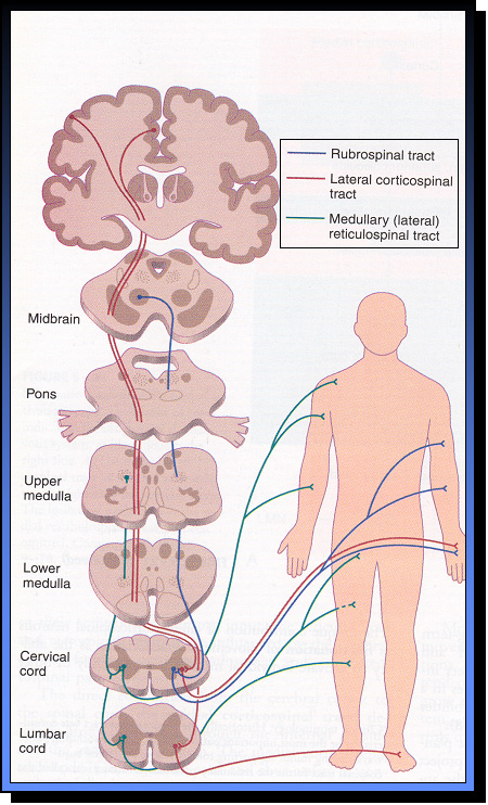
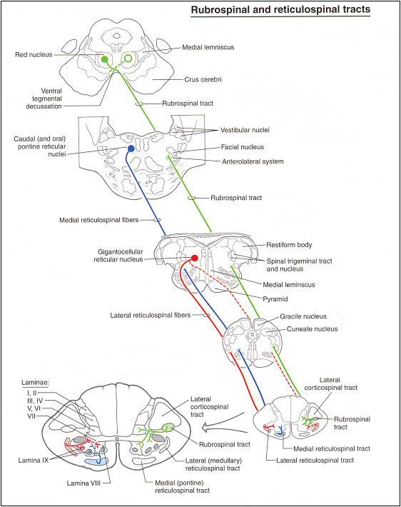
Rubrospinal Tract
Originates in the rd nucleus of the midbrain, crosses to opposite, then descends through the pons, medulla, and lateral spinal cord to synapse with lower motor neuron. Primarily innervating upper limb flexor muscles
-
Function :
- Activate flexor muscle
- Inhibit extensor muscle
Lateral(medullary) Reticulospinal Tract
-
Descends bilaterally
- to facilitate flexor muscle motor neurons
- to inhibit extensor motor neurons
-
Function :
Control alpha & gamma motor neuron
How Do each of the different descending tracts affect muscle tone ?
Each of the descending pathways has different influence on the background tone and dynamic activaction of motor neuron pools and interneuronal circuits in the spinal cord
The vestibulospinal and reticulospinal tracts are involyed in the postural biasing of muscle and anticipatory postual adjustments that precede voluntary novements
The vestibulospinal and reticulospinal output neuronsare
- generally excitatory to extensor motor neurons innervating extensor muscles in the arm and legs
- under inhibitory controal from the cortical level
Thus, loss of cortical inhibitory control over these pathways
- tend to facilitate extensor tone in the arms and legs, resulting in Decerebrate Rigidity
The robrospinal and corticospinal tracts both then to balance the extensor drive by facilitating drive to flexor muscles The rubrospinal tract in humans...
- extends only into the cervical cord
- thus can counteract extensor drive in the arm but not the legs.
Thus.... In humans....
Decorticate Rigldity is ... - primarily one of descending control from the cerebral cortex release unopposed excitutary extensor drive from the vestibular and reticular formation areas to the lower limb extensor muscles, while flexor facilitation is released from the rednucleus to upper limb flexor muscles in projections in the rubrospinal tract